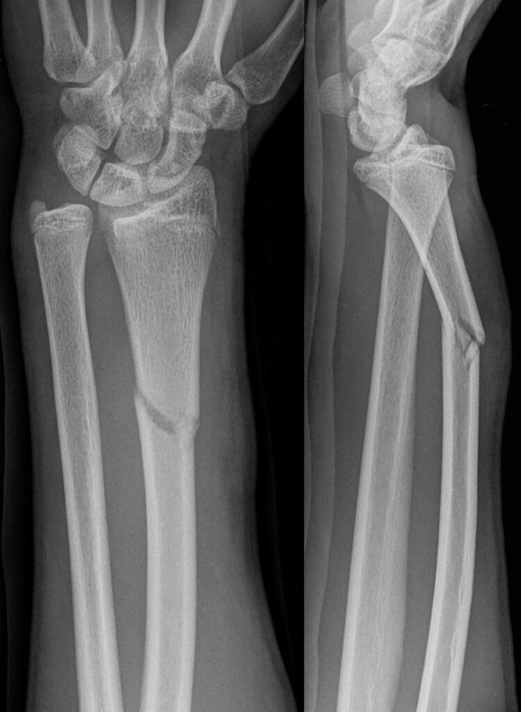
- Galeazzi fracture dislocation is a fracture of the middle to distal radius associated with a subluxation or dislocation of the distal radioulnar joint
- Galeazzi fractures are much more common than Monteggia fractures, Galeazzi account for 7% of all forearm fractures in adults
- Radius fractures occurring closer to the wrist are associated with greater instability of the DRUJ
Mechanism of injury
- Caused by a fall on the outstretched hand with extended wrist and pronated forearm and a rotational force applied to the body
- They also could occur from sports injuries and motor vehicle accidents
Clinical features
- Symptoms
- Patient present with pain in the forearm and wrist
- Physical examination
- Look
- Look for Swelling, contusions and lacerations
- Deformity of the radius or wrist might be obvious
- Inspect the lacerations for any evidence of open fracture
- Feel
- Feel for tenderness over the forearm and wrist
- Neurovascular examination is done to look for nerve/vessel injury (esp. median and radial nerves), inquire about weakness, numbness, paresthesia and motor examination
- Move
- Patient refuse movement due to pain
- Look
- Examination is repeated multiple times to exclude compartment syndrome
Imaging
- X-ray radiographs (forearm AP and lateral) should be ordered and they are enough for diagnosis
- A transverse or oblique fracture is seen in the radius with angulation or shortening,
- DRUJ is disrupted, signs include:
- Widening of DRUJ on AP
- Dorsal displacement of the ulna on the lateral view
- Ulnar styloid fracture
- Radial shortening greater than 5 mm

Note
- The distal radioulnar joint could be injured with isolated radial fracture at any level, or in both forearm bones fractures
Emergency management
- Pain management
- Gross forearm deformity should be reduced in the emergency department under procedural sedation
- Above elbow backslab is applied to support the fracture and prevent rotation
- Reassessment of neurovascular status is done
Definitive management
- Same as Monteggia, it is important to restore the length of the broken bone
- In children, closed reduction is often successful, but in adults closed reduction lead to poor outcomes
- That is why in adults, reduction is best achieved by ORIF and plating of the radius and the DRUJ is re examined and re imaged to ensure it is reduced
- If the DRUJ is reduced and stable on full range of movement, no further management is needed
- If the DRUJ is reduced but unstable then the forearm should be immobilized in the position of stability for the DRUJ (usually supination); K wires maybe applied to support the joint
- If the DRUJ is irreducible by closed reduction then open reduction is needed to remove soft tissue interposition or fracture fragment preventing reduction; the triangular fibrocartilage complex and dorsal capsule are then carefully repaired, if there is associated ulnar styloid fracture then it has to be repaired by ORIF
- After reduction in all cases, above elbow casting in supination is done for 6 weeks , exercises started as soon as possible
Complications
- Early
- Vascular injury
- Nerve injury
- Compartment syndrome: especially in high energy injuries
- Muscle tendon entrapment: make reduction difficult
- Late
- Malunion
- Non union
- Radioulnar synostosis
- Elbow stiffness
Course Menu
This article is apart from The Elbow and Forearm Trauma Free Course; This course contains a number of lectures listed below:
