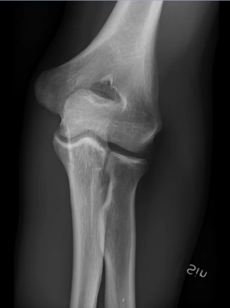
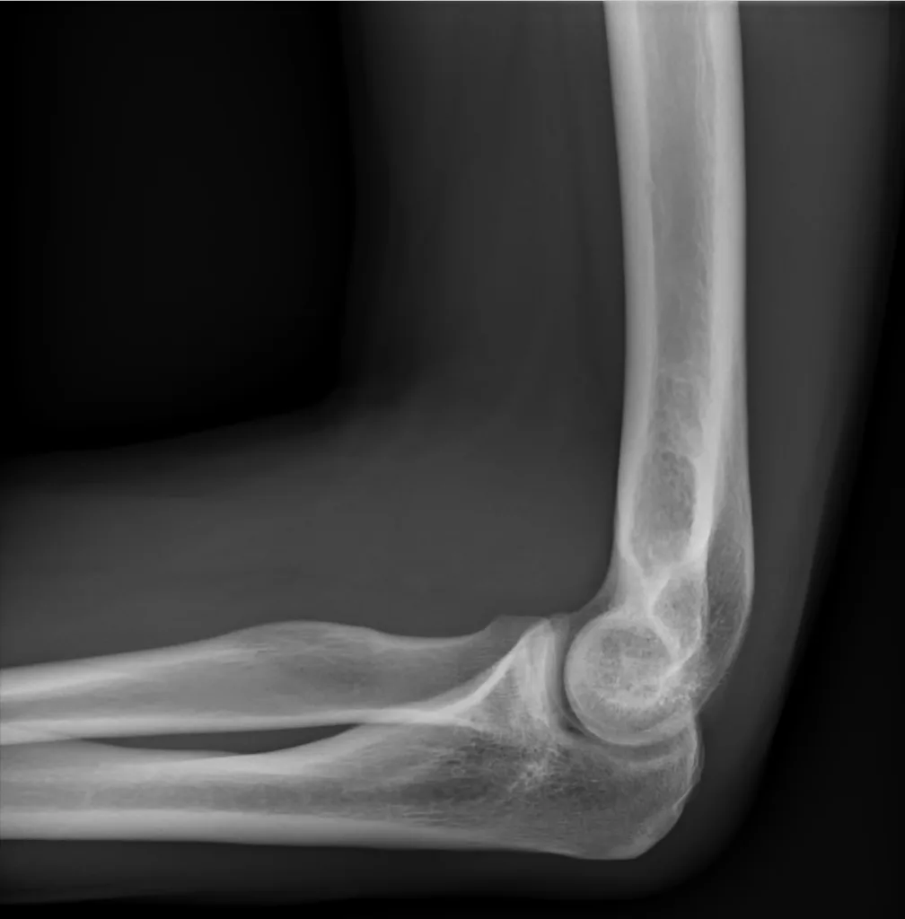
- Elbow X-ray interpreted by following this 3 steps approach:
- Step 1: assess Bones
- Step 2: assess Joints
- Step 3: assess Soft tissue
Step 1: assess Bones
- Trace the edges of each bone looking for cracks
- If you see the fracture then start with the fracture then look elsewhere
- Look at the general appearance of the bone looking for decrease or increase in density (osteopenia vs osteosclerosis) or abnormal trabeculation (Paget’s disease)


Step 2: assess Joints
- Assess the radiohumeral joint, ulnohumeral joint and radioulnar joint
- Look for the shape of the joint, congruity of the bone ends and look for narrowing or asymmetry
- Elbow lines: radiocapitellar line, anterior humeral line
Radiocapitellar line
- Radiocapitellar line is used to look for radial head dislocation
- Radiocapitellar line is drawn from the neck of the radius to the capitellum and should intersect the capitellum in all views

Anterior humeral line
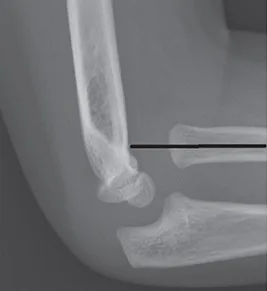
- Anterior humeral line is used to check for subtle supracondylar fracture on lateral elbow x-rays
- Anterior humeral line is drawn down the anterior surface of the humerus and should intersect the middle third of the capitellum
- In normal children younger than 4 years, anterior humeral line might intersect the anterior third of the capitellum and this is normal in this age group
Step 3: assess Soft tissue
- Look for changes in density, abnormal bulges, swelling, foreign bodies
- Higher density soft tissues are muscles while lower density soft tissues are fat
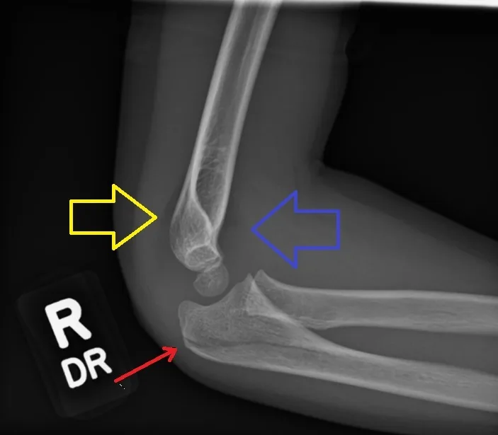
- On the lateral elbow radiograph, Inspect for elevation of the anterior and posterior fat pads; anterior fat pad can be seen in normal x-rays but posterior is not
- Elevation of these fat pads known as the sail sign, this indicates a joint effusion and it occur in elbow trauma, fractures and inflammation

Pediatric elbow X-rays
- Pediatric elbow X-rays is harder to be interpreted because of the ossification centers order of appearance
- The order of appearance for the elbow ossification centers: “CRITOE”
- Capitellum: 1 year of age
- Radial head: 3 years
- Internal epicondyle (medial): 5 years
- Trochlea: 7 years
- Olecranon: 9 years
- External epicondyle (lateral) : 11 years
Elbow X-ray views
- Primary radiographic series for elbow injuries is the AP and lateral views; those are the most common ones to be ordered
- Those are indicated in elbow injuries and various elbow conditions
- But there is other views that can be taken to look for specific elbow injuries or taken because of lack of patient mobility
- Those include:
- Modified trauma views
- Horixontal beam lateral:
- Acute flexion AP:
- Inferosuperior view
- Additional views
- Coyle’s view
- External oblique
- Inferior oblique view:
- Supraconylar AP
- Modified trauma views
Course Menu
This article is apart from The Elbow and Forearm Trauma Free Course; This course contains a number of lectures listed below:
