
- Distal radius fractures are common fractures that affect the distal radius
- They occur when a significant force is applied to the distal radius and this usually result from falls on the outstretched hand
- Distal radius fractures treatment include non operative and operative methods
Anatomy
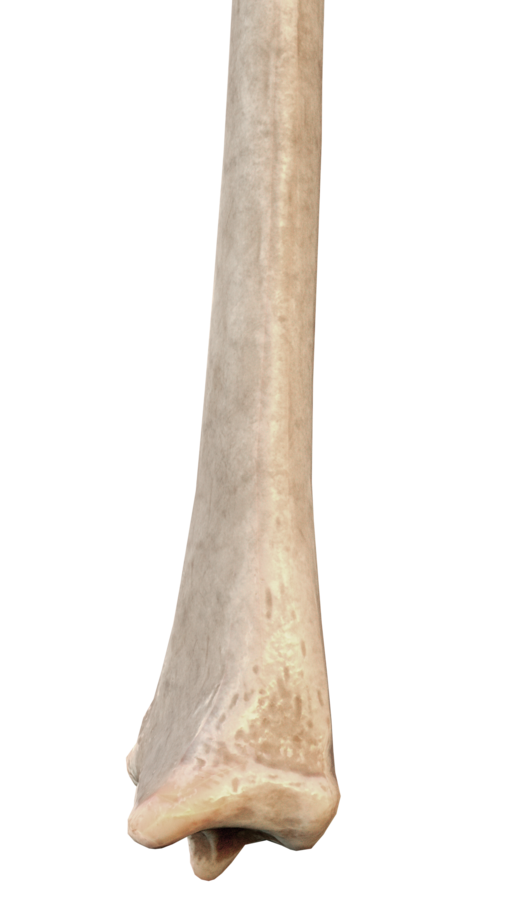
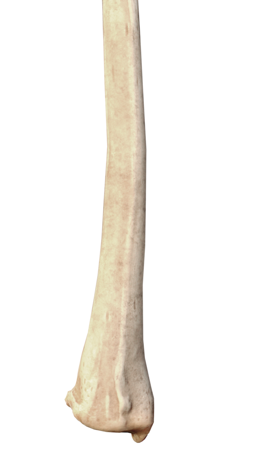
- the radial shaft enlarges as it moves distally (opposite to the ulna)
- The distal end of the radius has four different sides, its medial side forms a concavity (the ulnar notch) that accommodates the ulnar head
- Its lateral side forms a ridge terminating in the radial styloid process which is larger than the ulnar styloid process
- Posteriorly we have the dorsal tubercle of the radius which lies between the shallow grooves for the passage of the tendons of the forearm muscles

Classification
- Distal radius fractures are classified according to the fracture type, location, displacement, joint involvement and associated injuries using radiological assessment
- Eponyms names were given to the common fracture types of the radius: e.g. Colles fracture , Smith fracture …
- Although there are approximately 15 different classification systems (e.g. Frykman, AO …) but none appear to be clinically useful; so the normal radiological description and eponyms are used more to describe these fractures
- There are multiple factors that increase the severity of the distal radius fractures, those include:
- oblique, spiral, and comminuted fractures
- Intra articular fractures
- Significant displacement
- Associated fractures: distal ulnar fracture , ulnar styloid fracture, or scaphoid fractures
- Associated dislocation (e.g. DRUJ dislocation)
- Associated ligamentous injury: e.g. scapholunate dissociation
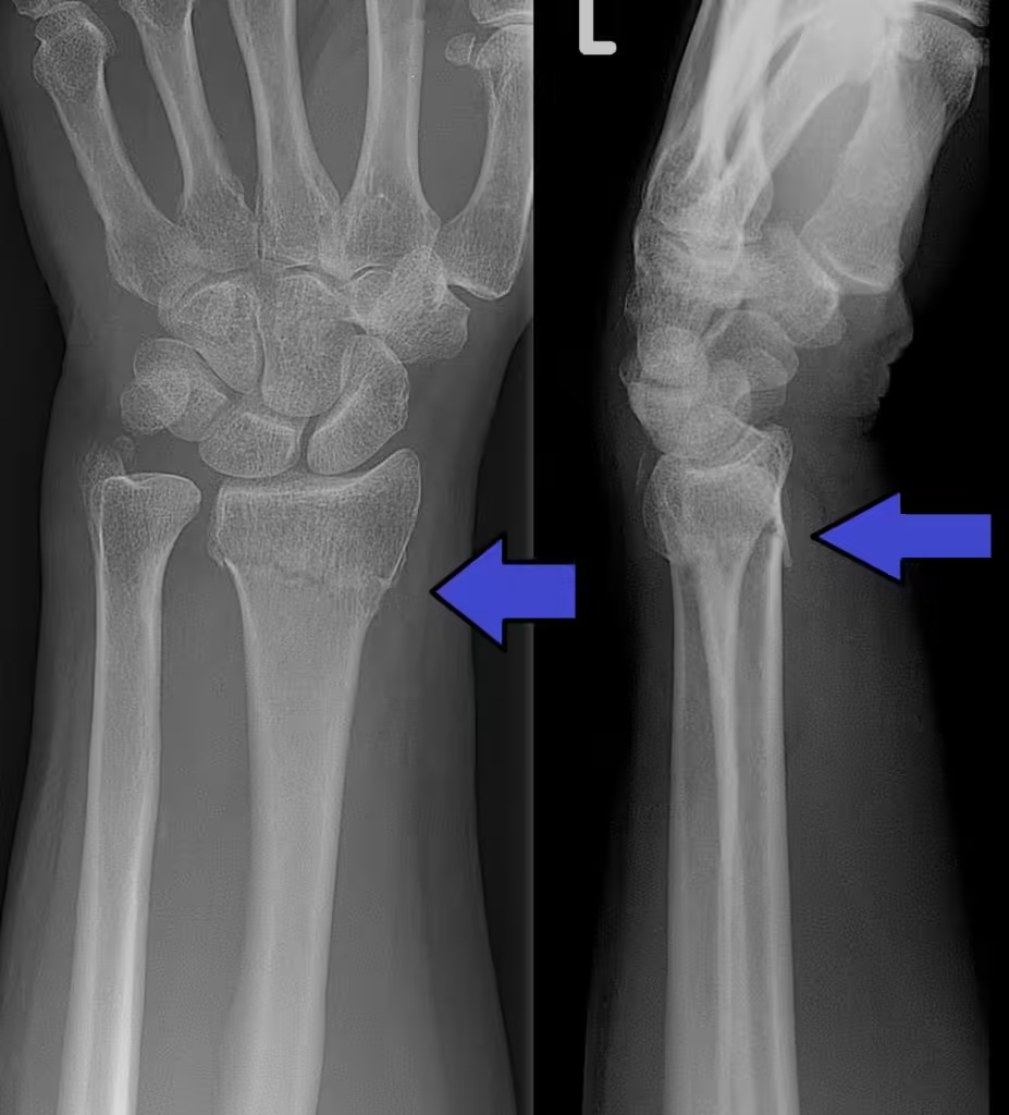
Colles fracture
- Colles fracture is an extra articular fracture of the distal radius with dorsal displacement
- Colles fracture is one of the most common fractures to affect the human body
- It mainly affect elderly patients after falling on the outstretched hand
- Ulnar styloid fracture is present in 50% of all Colles fracture cases
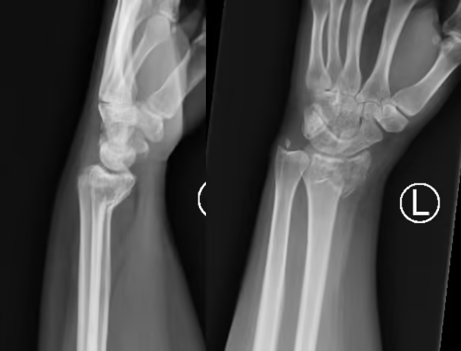
Smith’s fracture
- This is an oblique or a transverse extra articular fracture of the distal radius with volar displacement
- It is inherently unstable fracture and require surgical treatment
Barton’s fracture
- This is an oblique intra articular fracture of the distal radius with dorsal displacement (Dorsal Barton’s) or volar displacement (Volar Barton’s)
- This is an inherently unstable fracture
- It is associated with dislocation or subluxation of the radiocarpal joint
Chauffeur fracture
- This is an isolated intra articular radial styloid fracture caused by loading force on the scaphoid against the radius
- This results from starting handle of a car kicking back against the chauffeur’s hand ; same mechanism may result in a scaphoid fracture


Lunate fossa fracture
- Fracture of the volar aspect of the lunate fossa
- Lunate fossa provides attachment of the extrinsic carpal ligaments, displacement of this fragment may result in subluxation of the entire carpus
Die punch fragments
- Intraarticular fragmented fracture with displacement in form of a step or a gap (Die-punch) that occur due to axial loading force
- A section of the articular surface impacted below the level of the adjacent cartilage
- These fractures may require delicate manipulation and fixation with levers and wires
Clinical features
Symptoms
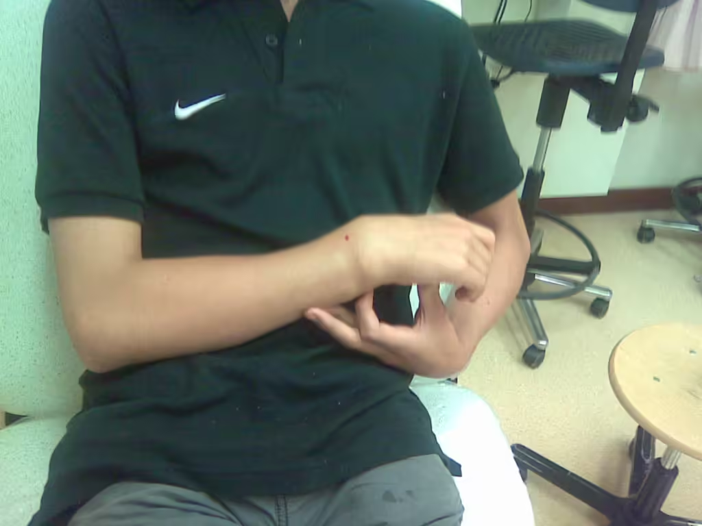
- Patient present tucking their wrist to the chest and supporting it with the other hand and they complain of wrist pain
Physical examination
- Look
- Look for Swelling, contusions and lacerations
- Deformity maybe clear; Colles’ fracture is associated with dinner fork deformity if there is significant displacement.
- Inspect the lacerations for any evidence of open fracture
- Feel
- Feel for tenderness over the forearm, wrist and hand
- Neurovascular examination is done to look for nerve/vessel injury, inquire about weakness, numbness, paresthesia and motor examination
- Move
- Patient refuse movement due to pain
- Examination is repeated multiple times to exclude compartment syndrome
Imaging
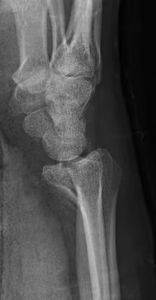
- AP and lateral wrist radiographs are usually enough to make the diagnosis
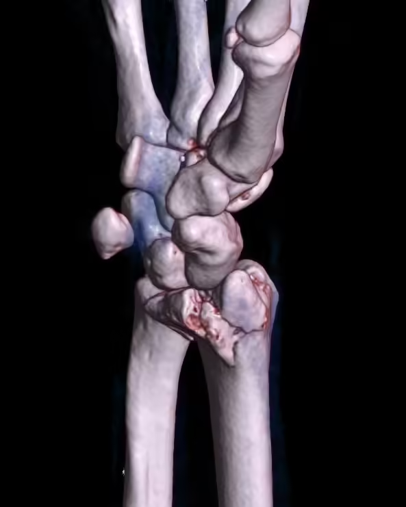
- CT scan is indicated in highly comminuted fractures and for preoperative planning
- MRI is helpful for occult fractures and for ligamentous injuries
- When describing a distal radius fracture; comment on:
- Type of fracture
- Displacement
- Joint involvement
- Associated fractures
Treatment
- Non operative
- Operative
Non operative treatment
- Most distal radius fractures are treated non operatively
- Minimally displaced or non displaced fractures are immobilized with a cast for 4-6 weeks or a splint and supported with a sling, the wrist will be slightly flexed with ulnar deviation inside the cast
- Fractures with significant displacement will require closed reduction through traction and manipulation and then immobilization with a cast is done for 4- 6 weeks
- Neurovascular examination is done again after the application of the cast to make sure no further injuries occurred during reduction or cast application
- X-ray radiographs are ordered again after the application of the cast to make sure the reduction is good
- Exercises are started as soon as pain allows
Operative treatment
- Operative treatment is done through percutaneous wires or ORIF with plating or intramedullary nails
- Indications
- Comminuted fractures
- Unstable fractures: fracture re displaces after reduction, fracture with volar displacement (all smith’s and volar Barton’s are considered unstable)
- Intra articular fractures with > 2mm displacement
- Open fractures
- Un reducible fractures
- Die punch fracture
Complications
- Early
- Vascular injury
- Nerve injury: median nerve can be damaged during the injury or during the operative treatment and if the symptoms are mild this might be resolved with release of the dressing and elevation but if the symptoms are severe then this suggest acute carpal tunnel syndrome which require urgent intervention through dividing the transverse ligament
- Redisplacement: that is why X-rays are ordered again after 1 week
- Compartment syndrome: especially in high energy injuries
- Extensor pollicis longus tendon rupture
- circulatory problems due to fingers venous circulation not draining well leading to finger cyanosis, that is why fingers must be checked regularly and elevation and finger movements is done to prevent that
- Late
- Malunion: fracture unite at abnormal anatomical position
- Post traumatic carpal tunnel syndrome
- Secondary osteoarthritis: especially in patients with intra articular involvement
- CRPS: common after these injuries, swelling and tenderness of finger joints and vasomotor changes such as altered sweating