Understanding Bone healing process is important in treatment of fractures in many ways that will be explained here, it is also helpful in expecting the time by which the fracture heals.
Requirements for fracture healing
First we need to know what is needed for the fracture to heal, this include:
- Intact blood supply to the fractured fragments
- Immobilization by cast, internal fixation or external fixation
- Absence of infection
Bone healing types
We have three types of bone healing
- Secondary bone healing (indirect, spontaneous)
- Primary bone healing (contact healing)
- Gap healing
If there is small amount of tension (below 2%) on the fracture (the fracture ends held very stable) => primary bone healing occurs; such as when using plates and screws (type of internal fixation)
If the tension is between 2-10 % (there is a little movement between the fracture ends due to external forces) => secondary bone healing will occur such as when using a cast, intramedullary nail or external fixation
1. Secondary bone healing (healing by callus)
Secondary bone healing is on 5 stages:
- Stage one: Hematoma formation
- Stage two: Inflammation
- Stage three: Soft callus formation
- Stage four: Hard callus formation
- Stage five: Remodeling
Stage one: Hematoma formation
- Initially when the fracture occur, it would lead to bleeding coming from the bone and soft tissue at the fracture site
- This ultimately lead to collection of blood known as hematoma

Stage two: Inflammation
- After the hematoma forms, inflammatory process start
- Cytokines are released
- Inflammatory cells recruited (neutrophils, macrophages, monocytes, T cells)
- Osteoclasts formed to remove necrotic ends of bone fragments
- Last until fibrous tissue, cartilage or bone formation begins (1-7 days) after fracture

Stage three: Soft callus formation
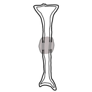
- Callus is a temporary bony material, cartilaginous material, fibrous connective tissue and woven bone that replaces the hematoma
- Woven bone is bone tissue characterized by haphazard arrangement of collagen fibers and will be remodeled into cortical bone in the upcoming stages
- Soft callus: soft bony material (collagen type 2= cartilage) so it is mainly formed by cartilage, secreted by mesenchymal stem cells that are gathered to injury site because of the inflammation process
- Soft callus forms After 2 weeks post fracture
- The fragments can’t move freely now, they are stable in length but they can angulate
- The stiffer the immobilization of the fracture the less amount of callus formed

Mesenchymal stem cells
- Mesenchymal stem cells are undifferentiated and they differentiate to osteoblasts and chondroblasts
- Important sources of mesenchymal stem cells in fractures include periosteum, bone marrow and surrounding muscles
- Gathered during the inflammation process
Stage four: Hard callus formation
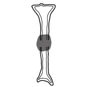
- Hard callus: hard bony material (woven bone which is type 1 collagen) so it is mainly bone material, secreted by osteoblasts and chondroblasts
- Starts forming after the formation of the soft callus and ends when the fragments are firmly united by 3-4 months post fracture
- Forms in the periphery of the fracture and progressively moves centrally

Stage five: Remodeling
- The woven bone is slowly replaced by lamellar bone
- The medullary bone canal is continuous in this stage
- Last from few months to several years post fracture

2. Primary bone healing (contact healing)
- Bone ends are put in close contact
- When fracture surfaces are in intimate contact (less than 0.1 mm distance) with absolute stability => osteoclasts will form cutting cones that traverse the fracture line => capillaries occupy the newly formed cavities with osteoblasts => form lamellar bone from osteons
- The intermediate stages are skipped (no callus what so ever)
- Primary bone healing process called cutting cone remodeling, Haversian remodeling
3. Gap healing
Gap healing occurs when there is small gaps between fracture surfaces (< 1mm) => gaps invaded by new capillaries and osteoblast cells that form => woven bone => remodeled to lamellar bone
Factors affecting fracture healing
- Age of the patient: healing is faster in children, the time to heal a fracture in children is half compared to adults
- Type of bone: cancellous bones unite faster than cortical bones
- Fracture pattern: spiral heals faster than > transverse > comminuted
- Ischemic fracture ends (due to damage to the blood supply from trauma or from fixation) lead to slower healing or no healing at all
- Soft tissue interposition between fracture ends prevent callus from bridging the fragments and lead to slower healing
- Reduction: good apposition of the fracture ends lead to faster healing
- amount of movement at fracture site (movement lead to callus formation accordingly):
- Absolute stability and compression leads to primary bone healing
- Relative stability leads to secondary bone healing
- Excessive motion leads to delayed healing or non union
Average healing times of some fractures in adult patients
- Upper limb
- Clavicle, humerus, radius, ulna: 6-8 weeks
- Wrist bones: 6-12 weeks
- Hand/ fingers: 4-6 weeks
- Lower limb
- Hip, femur, tibia, fibula: 6- 12 weeks
- Ankle bones: 6-8 weeks Foot bones: 4-6 weeks
Average healing times of fractures in pediatric patients
- 4-6 weeks in most fracture
Course Menu
This article is apart from Orthopedic Trauma Basic Principles Course, This course covers these topics:
